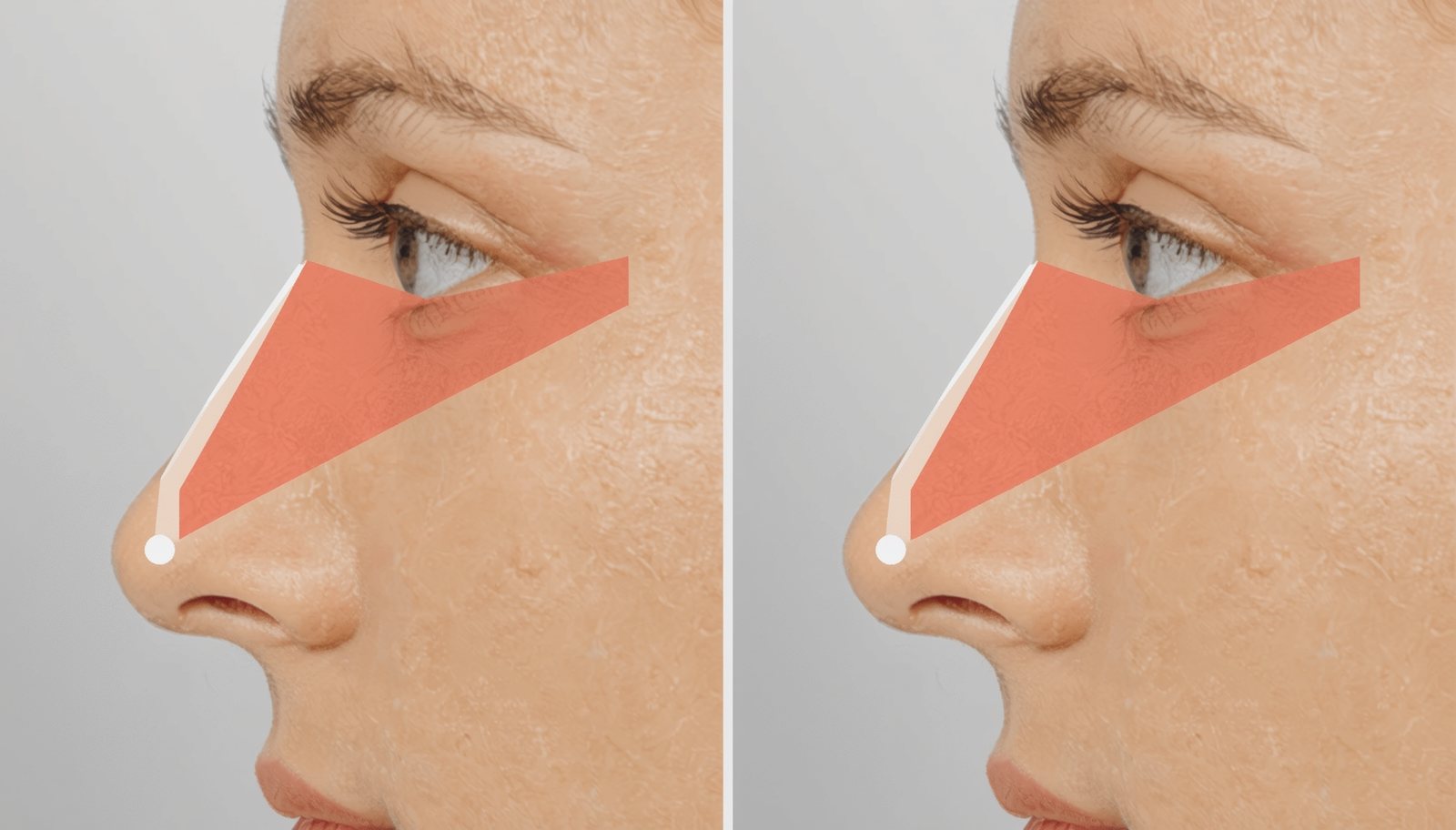
The Double-Edged Sword of Thin Skin
Thin nasal skin presents both tremendous opportunity and significant challenge in rhinoplasty. Measuring just 2-3mm thick, this delicate, often translucent envelope beautifully reveals refined contours and precise tip definition—the hallmark of elegant rhinoplasty results. However, this same transparency mercilessly exposes every minor asymmetry, irregularity, or technical imperfection, demanding near-perfect surgical execution and meticulous attention to detail.
Patients with thin skin often describe their nose as having a parchment-like quality where underlying structures are visible, particularly in bright lighting. The skin appears smooth, poreless, and sometimes slightly shiny, with minimal sebaceous (oil gland) activity. This skin type is common among individuals of Northern European, Persian, and some East Asian descent, though it occurs across all ethnic backgrounds.
The translucency that makes thin skin rhinoplasty so rewarding also means that post-surgical refinement continues for years as the shrink-wrap effect progressively tightens the skin against the underlying framework, sometimes revealing structures or creating contours not immediately apparent after surgery.
Anatomical Characteristics of Thin Skin
Thin nasal skin differs from thick skin not just in millimeter measurements but in fundamental tissue composition and behavior. The dermis contains fewer collagen and elastin fibers, creating less structural bulk but also less elasticity. The subcutaneous layer contains minimal adipose (fat) tissue, meaning skin sits almost directly on cartilage and bone with little cushioning buffer.
Sebaceous glands are sparse and small, producing minimal oil and creating the characteristic smooth, matte appearance. This lack of oil activity means thin skin generally responds poorly to isotretinoin (Accutane) protocols that work well for thick, sebaceous skin, as there's minimal sebaceous activity to suppress.
The vascular network, while present, is more delicate than in thick skin. This requires careful surgical technique to preserve blood supply, as thin skin is more vulnerable to vascular compromise, though actual skin necrosis remains rare with proper technique. The rich superficial capillary plexus often creates a pinkish hue, particularly visible in very fair-skinned individuals.
Importantly, thin skin has remarkable contractile properties—the shrink-wrap effect that causes progressive tightening over 2-3 years post-surgery. This contraction can be both blessing and curse: it beautifully reveals refined contours but can also accentuate minor asymmetries or irregularities not apparent immediately after surgery.
The Shrink-Wrap Effect: Years of Evolution
One of the most distinctive features of thin skin rhinoplasty is the prolonged evolution of results through the shrink-wrap phenomenon. Unlike thick skin that maintains relatively stable contours once swelling resolves, thin skin continues contracting and conforming to the underlying framework for 2-3 years or even longer.
In the early post-operative period (1-6 months), thin skin patients often see beautiful initial results with minimal swelling obscuring refined contours. However, as months progress into years, the skin continues tightening, sometimes revealing structures that weren't visible initially. A cartilage graft that was perfectly smooth under swollen skin at 3 months might develop visible edges at 18 months as skin contracts further.
This phenomenon demands surgical foresight—anticipating how structures will appear not just after swelling resolves but after years of skin contraction. Grafts must be meticulously smoothed and beveled. Suture techniques must create soft, natural contours rather than sharp angles. Any asymmetry must be corrected to the point of near-imperceptibility, knowing thin skin will eventually reveal what thick skin would forever hide.
The positive aspect of this effect is the emergence of increasingly refined definition over time. Patients often report their nose looks even better at 2-3 years than it did at 6-12 months as the skin molds ever more precisely to the refined framework, creating exquisite tip definition and elegant lines impossible with thicker skin types.
Surgical Philosophy: Perfection as Standard
The fundamental principle of thin skin rhinoplasty is simple but demanding: near-perfect execution is the minimum acceptable standard. There is no margin for error, no close enough, no relying on thick skin to camouflage minor imperfections. Every structure must be precisely symmetric, every graft perfectly smooth, every edge meticulously beveled, and every suture placed with attention to creating soft, natural contours.
This perfectionistic approach extends through every aspect of surgery. Dorsal reduction must create glass-smooth surfaces without step-offs or irregularities. Tip work must achieve symmetric domes with gentle curves and natural light reflexes. Any grafts must be carved, thinned, and placed with microscopic precision. The underlying framework must be as flawless as a sculpture meant for display without covering.
Surgeons experienced in thin skin rhinoplasty develop obsessive attention to detail, often spending significantly more time smoothing edges, checking symmetry, and refining contours than they would in thick skin cases where these nuances would remain invisible. The surgical magnification (loupes or microscope) often used becomes essential for this level of precision.
Dorsal Management: Glass-Smooth Perfection
The nasal dorsum (bridge) in thin skin rhinoplasty must achieve absolutely smooth contours from radix (root) to tip. Any irregularity—a small step-off after hump removal, a slightly asymmetric osteotomy line, a raised suture knot—will eventually become visible as skin contracts.
When dorsal reduction is performed, surgeons must remove the hump with extraordinary precision, ensuring the cartilaginous and bony reductions blend seamlessly. Traditional techniques using rasps to smooth bone must be executed meticulously, as even micro-irregularities can become palpable or visible through thin skin. Some surgeons prefer powered instrumentation (high-speed burrs) for bone work in thin skin patients, as these create smoother surfaces than manual rasps.
The open roof deformity created after hump removal must be closed with precisely executed osteotomies. The nasal bones must be mobilized and positioned symmetrically, with smooth contours and no palpable steps where bone edges meet. In thin skin patients, even 1mm asymmetry in osteotomy position can create visible shadowing or light reflection differences.
When dorsal augmentation is needed—building height rather than reducing it—thin skin presents different challenges. The augmentation material (typically cartilage) must be precisely carved and surfaced to create natural contours. Multiple thin layers often work better than single thick grafts, as they can be stacked and shaped more precisely.
Specialized techniques for thin skin dorsal work include diced cartilage wrapped in fascia (DC-F technique), which creates soft, moldable augmentation that won't show sharp edges, precise cap grafts beveled at edges to create imperceptible transitions, and camouflage grafts that smooth minor irregularities by bridging rather than filling defects.
Tip Refinement: Sculpting for Transparency
The nasal tip represents both the greatest opportunity and highest risk in thin skin rhinoplasty. Thin skin beautifully reveals refined tip definition, elegant tip-defining points, and sophisticated light reflexes—the hallmarks of artful rhinoplasty. However, any asymmetry, irregularity, or technical imperfection becomes glaringly obvious.
Suture techniques form the foundation of thin skin tip work. Interdomal sutures bring tip-defining points (domes) closer together, narrowing the tip. Transdomal sutures create more acute dome angles, increasing definition. These sutures must be placed with precision, creating symmetric, natural contours rather than pinched or overly narrow tips that look operated.
The tension applied to these sutures is critical in thin skin patients. Excessive tension can create unnatural sharpness or visible dimpling of the skin. Insufficient tension produces inadequate refinement. The suture material itself matters—many surgeons prefer clear (polypropylene) permanent sutures that won't show through thin skin like colored sutures might.
When grafts are needed for tip refinement—shield grafts, cap grafts, or tip onlay grafts—they must be carved from the finest cartilage available (often septum) rather than thicker conchal or costal cartilage. The graft must be thin enough to avoid bulk but substantial enough to create desired change, typically 1-2mm thick compared to 3-4mm used in thick skin cases.
Every graft edge must be beveled (tapered) so the transition from graft to native cartilage is imperceptible. Some surgeons pre-thin grafts by crushing or morselizing edges, creating gradual rather than abrupt thickness changes. The graft must be positioned and secured so it won't shift or rotate during healing, as even subtle displacement becomes visible through thin skin.
Dealing with Visible Irregularities: Camouflage Techniques
Despite meticulous technique, thin skin sometimes reveals minor irregularities from the underlying framework—a slightly visible graft edge, a small cartilage asymmetry, a palpable suture. Specialized camouflage techniques help smooth these issues without major structural revision.
Diced cartilage techniques have revolutionized thin skin rhinoplasty. Cartilage is finely diced (0.5-1mm pieces) and wrapped in temporalis fascia or another tissue envelope. This creates a soft, moldable graft that can be placed over irregularities, smoothing contours without creating additional edges or irregularities. The diced cartilage integrates with surrounding tissue, maintaining volume while the fascial wrap creates a smooth surface that won't show through thin skin.
Fascia lata grafts—thin sheets of fascia harvested from the thigh—provide another excellent camouflage material. These paper-thin grafts can be layered over the dorsum or tip to smooth irregularities without adding significant bulk. The fascia integrates with native tissue and creates an additional buffering layer between skin and underlying structures.
Perichondrium flaps—the thin membrane covering cartilage—can sometimes be elevated, extended, and draped to camouflage edges without requiring donor site harvest. This autogenous tissue integrates perfectly and adds minimal bulk while providing crucial camouflage.
Dermofat grafts represent another option for camouflaging and soft tissue augmentation in thin skin patients, particularly for revision cases with multiple irregularities. These composite grafts (dermis with attached subcutaneous fat) provide both structural fill and surface smoothing.
Conservative Approaches: Less is More
A fundamental principle in thin skin rhinoplasty is conservation—removing less tissue, making smaller modifications, and avoiding aggressive techniques that might look acceptable initially but create problems as skin contracts. The shrink-wrap effect means that conservative reduction at surgery may appear more aggressive after years of skin contracture.
Tip work in particular requires restraint. While thick skin patients may need aggressive structural enhancement to achieve visible change, thin skin patients can achieve dramatic refinement with subtle modifications. A 2mm dome-bringing suture might create more apparent tip narrowing than a 4mm suture in a thick-skinned patient, simply because the change is fully visible rather than obscured.
Cartilage preservation when possible—maintaining native structures rather than removing and replacing them with grafts—often produces the most natural results. The native lower lateral cartilages, when properly reshaped with sutures, create contours that integrate seamlessly with thin skin because they've evolved together. Even the finest grafts remain foreign structures that must integrate.
Spreader grafts in the middle vault should be thin (1-1.5mm) rather than thick, providing functional airway support without creating visible bulk along the dorsum. Columellar struts should be precisely carved to fill the space between medial crura without creating palpable or visible thickness.
The Open Approach: Seeing What You're Creating
While the open versus closed rhinoplasty debate continues, many surgeons favor the open approach specifically for thin skin patients because it provides complete visualization of the structures that will eventually be revealed through transparent skin. The open approach involves a small transcolumellar incision connecting to bilateral marginal incisions, allowing the nasal skin to be elevated and draped back, exposing the entire cartilaginous framework.
This complete exposure allows surgeons to assess symmetry with both eyes viewing from directly above, achieve meticulous graft placement with precise positioning, execute complex suture techniques with full visibility, and identify and correct minor irregularities that would be difficult to appreciate through endonasal approaches. The transcolumellar scar, when properly placed in the narrowest part of the columella and closed meticulously, becomes virtually invisible, even through thin skin.
Some surgeons still favor closed approaches for thin skin cases requiring minimal tip work, arguing that less dissection means faster healing and potentially less shrink-wrap effect. However, for complex thin skin rhinoplasty requiring multiple grafts, sophisticated suturing, or revision work, the open approach's advantages typically outweigh concerns about the small external incision.
Managing Patient Expectations: Slow Reveal
One unique aspect of thin skin rhinoplasty counseling involves preparing patients for the prolonged evolution of results. Unlike thick skin patients who must wait patiently for swelling to resolve before seeing definition, thin skin patients see beautiful initial results that then continue evolving—sometimes in unexpected ways—for years.
This requires explaining the shrink-wrap effect and its timeline, discussing that results at 6 months may differ from results at 2 years, managing anxiety if patients notice new contours or shadows emerging after the first year, and ensuring patients understand that this evolution is normal and expected, not a complication or surgical error.
Surgeons must also temper enthusiasm about rapid initial results, reminding patients that while they look great at 3 months, the final result won't be apparent for 2-3 years. This prevents patients from assuming they're done healing and becoming alarmed when contours continue changing.
Progressive photography—images at 3, 6, 12, 24, and 36 months—helps patients and surgeons track this evolution and appreciate subtle improvements or identify issues requiring intervention while they're still amenable to minimally invasive correction.
Revision Rhinoplasty Considerations
Thin skin patients face unique challenges in revision rhinoplasty. The skin has already undergone one shrink-wrap cycle and will undergo another. Scar tissue from the previous surgery creates irregularities that thin skin may reveal. Cartilage supply may be limited if septum and possibly ear cartilage were harvested previously.
Revision surgery in thin skin patients requires even more meticulous technique than primary surgery. Every previous irregularity must be addressed, scar tissue must be carefully released or excised, and grafting must create flawless contours knowing thin skin will reveal any imperfection. The use of rib cartilage becomes more common in thin skin revisions, as this abundant source allows creation of extensive structural grafts for comprehensive framework reconstruction.
Fascia grafts, diced cartilage techniques, and other camouflage methods become essential in thin skin revision cases where multiple irregularities from previous surgery must be smoothed. The surgeon must essentially create a new, perfect framework beneath skin that has already contracted and may have limited remaining elasticity.
Advanced Technologies for Thin Skin Planning
Modern imaging technologies particularly benefit thin skin rhinoplasty planning. High-frequency ultrasound allows preoperative measurement of actual skin thickness, identifying areas where skin is thinnest (often the rhinion—where bone meets cartilage—and the nasal tip), assessing underlying cartilage morphology, and detecting minor asymmetries that will become apparent post-operatively.
3D surface imaging captures precise preoperative contours, allows virtual surgical simulation showing potential outcomes, provides objective measurements for surgical planning, and creates comparison models for postoperative evaluation. This technology helps surgeons identify asymmetries that might not be obvious on 2D photographs and plan corrections that achieve true three-dimensional symmetry.
Intraoperative technologies including surgical loupes or microscopes for magnified visualization, high-definition endoscopy for endonasal assessment, and real-time imaging ensuring perfect graft placement and symmetry all contribute to the precision required for thin skin excellence.
Choosing Your Surgeon: Precision Expertise Required
The success of thin skin rhinoplasty depends absolutely on surgeon selection. Not all experienced rhinoplasty surgeons have equal facility with thin skin's unforgiving demands. During consultations, specifically ask about the surgeon's experience with thin skin patients, request before-and-after photos of patients with similar skin characteristics including long-term follow-up (2-3 years), inquire about techniques for achieving smooth contours and camouflaging potential irregularities, and discuss realistic expectations for initial results versus long-term evolution through shrink-wrap effect.
Look for surgeons who demonstrate obsessive attention to detail in their consultation, who spend time discussing nuances and potential challenges rather than offering overly simplistic or overly optimistic predictions, who show understanding of thin skin's unique characteristics rather than treating all rhinoplasty patients identically, and who have a portfolio demonstrating consistently refined, natural results in thin-skinned patients.
AI-powered matching platforms can filter surgeons by specific expertise in thin skin rhinoplasty, identifying those who consistently achieve elegant refinement with thin skin patients and whose technique emphasizes the precision thin skin demands.
Conclusion: Embracing the Canvas of Transparency
Thin skin rhinoplasty represents the pinnacle of surgical artistry—a specialty within a specialty that demands not just technical skill but perfectionism, foresight, and artistic sensibility. When executed with meticulous attention to detail by a surgeon who understands thin skin's unique characteristics, the results achieve a level of refinement and elegance simply impossible with thicker skin types. Your translucent canvas, while unforgiving of imperfection, beautifully reveals the sophisticated sculpting that defines exceptional rhinoplasty outcomes—results that grow even more refined and beautiful over years as your skin molds to the perfected framework beneath.