The Architectural Blueprint of Your Nose
Your nose is far more than a simple feature—it's a sophisticated three-dimensional structure composed of bone, cartilage, soft tissue, and skin working in perfect harmony. Understanding nasal anatomy is essential not only for surgeons but also for patients considering rhinoplasty, as it provides insight into what can realistically be achieved and why certain approaches work better for different nose types.
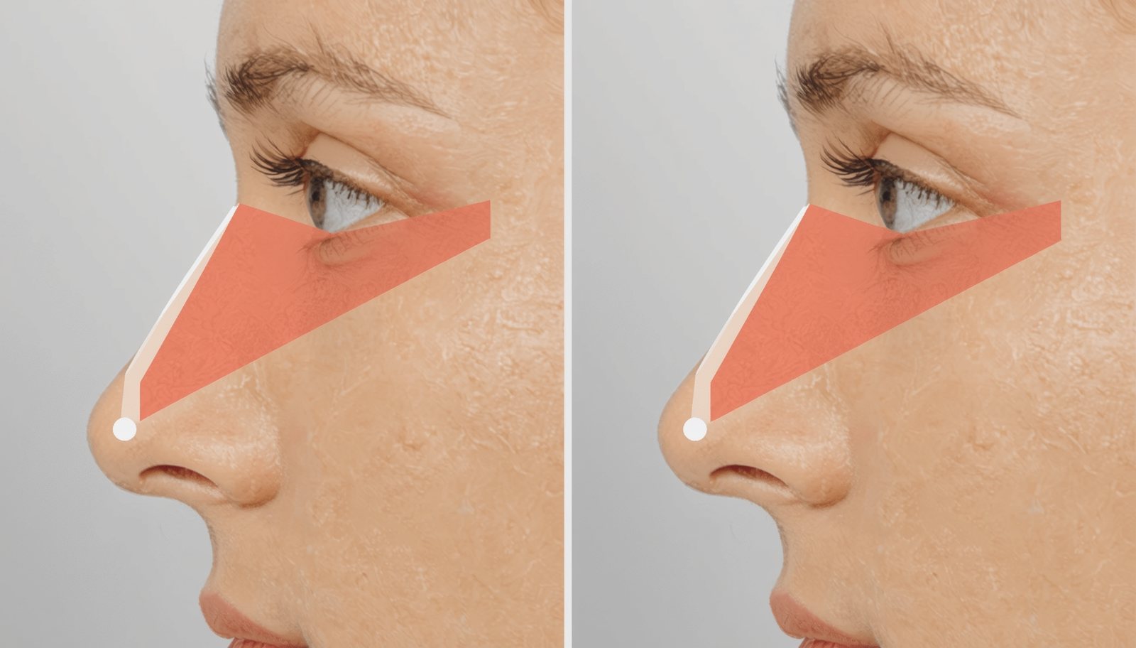
The nose consists of distinct anatomical zones, each playing a critical role in both appearance and function. From the rigid bony vault at the top to the flexible cartilaginous tip at the bottom, every component must be carefully considered during surgical planning.
The Bony Framework: Upper Third Foundation
The upper third of the nose comprises the nasal bones and portions of the maxilla and frontal bones. These paired nasal bones create the solid bridge that extends from between your eyebrows down to approximately the middle of your nose. The thickness and width of these bones vary significantly among individuals and ethnic backgrounds, affecting both the starting point for surgery and the techniques required.
The nasion marks the junction between the nasal bones and the frontal bone, creating the deepest point at the root of the nose. The rhinion represents the junction where bone transitions to cartilage—a critical landmark for surgical planning. Understanding these bony landmarks helps surgeons determine appropriate reduction amounts and the necessity for osteotomies (controlled bone fractures) to narrow or straighten the nasal bridge.
The Cartilaginous Middle Vault: Structural Support
The middle third of the nose features the upper lateral cartilages, which extend from beneath the nasal bones down toward the tip. These paired triangular cartilages attach to the nasal septum medially and overlap with the lower lateral cartilages inferiorly. This region houses the internal nasal valve—the narrowest part of the nasal airway and a common site of breathing obstruction.
The relationship between the upper lateral cartilages and the septum creates what surgeons call the keystone area, a critical structural junction that must be preserved or carefully reconstructed during dorsal hump reduction. Disruption of this area without proper reconstruction can lead to the inverted-V deformity or midvault collapse, causing both aesthetic concerns and breathing difficulties.
The Nasal Septum: The Central Pillar
The nasal septum is the wall dividing your nasal cavity into right and left chambers. It consists of several components: the quadrangular cartilage anteriorly, the perpendicular plate of the ethmoid bone superiorly, the vomer bone posteriorly, and the maxillary crest inferiorly. This complex structure provides crucial support for the entire nose.
During rhinoplasty and septoplasty procedures, surgeons typically preserve an L-strut of at least 10-15mm of dorsal and caudal septum to maintain structural integrity and prevent collapse. The septum also serves as an invaluable source of cartilage grafts, which can be used to build structure, provide support, or camouflage irregularities elsewhere in the nose.
Septal deviations—crookedness of this central structure—are extremely common and can result from developmental abnormalities, trauma, or previous surgery. Correcting these deviations often requires careful remodeling techniques including scoring (weakening), suturing, or replacement with straightened cartilage segments.
The Lower Lateral Cartilages: Defining the Tip
The nasal tip is shaped by the paired lower lateral cartilages (also called alar cartilages), which consist of medial crura, middle crura (domes), and lateral crura. The domes—the highest points of these cartilages—largely determine tip definition and shape. The space between the domes creates the tip's width, while their orientation affects tip rotation and projection.
The strength, thickness, and resilience of these cartilages vary dramatically between individuals. Strong, thick cartilages provide excellent structural support but can be resistant to reshaping, while weak cartilages may require reinforcement with grafts to achieve desired definition. Ethnic variations are particularly notable here, with some populations having naturally more robust or more delicate tip cartilages.
The medial crura form the columella (the external strip between the nostrils), while the lateral crura extend into the nasal sidewalls. Structural grafts like columellar struts, tip grafts, and alar rim grafts work with these native structures to achieve refined yet stable results.
Skin Types and Their Impact on Surgical Outcomes
Perhaps no factor influences rhinoplasty results more profoundly than skin characteristics. Nasal skin varies in thickness, elasticity, sebaceous (oil gland) activity, and capacity to contract or re-drape over the modified framework beneath.
Thin skin (2-3mm thick) offers maximum definition and detail visibility. Every subtle refinement shows through beautifully, but so does every minor imperfection. Surgeons must achieve near-perfect symmetry and smoothness, as thin skin provides no camouflage for irregularities. The shrink-wrap effect—progressive skin contraction during healing—is more pronounced with thin skin, sometimes revealing grafts or causing asymmetries months after surgery.
Thick skin (4-6mm or more) presents the opposite challenge. The heavy skin envelope obscures fine details and requires more aggressive structural modifications to achieve visible refinement. Thick-skinned patients must understand that their results will emphasize overall shape and proportion rather than fine tip definition. However, thick skin provides excellent camouflage for minor irregularities and typically produces very natural-looking results. Swelling resolution takes considerably longer—sometimes 2-3 years for complete definition to emerge.
Sebaceous (oily) skin requires special consideration, as the abundant oil glands contribute to thickness and may respond to treatments like low-dose isotretinoin (Accutane) to reduce sebaceous activity and improve skin draping. Recent advances include preoperative skin conditioning protocols to optimize surgical outcomes in thick-skinned patients.
Nasal Ligaments and Soft Tissue Envelope
Modern preservation rhinoplasty techniques have renewed focus on the nasal soft tissue envelope and its intricate ligamentous attachments. The scroll ligament connects upper and lower lateral cartilages, while the Pitanguy ligament connects the dermis to underlying cartilage in the supratip region.
These structures help maintain natural contours and anatomical relationships. Preservation techniques aim to maintain these connections when possible, reducing postoperative swelling, improving skin redraping, and creating more natural, stable long-term results compared to traditional approaches that disrupted these attachments.
Blood Supply and Innervation
The nose receives rich blood supply from both the internal and external carotid systems. The external nasal artery, angular artery, and dorsal nasal artery create an extensive vascular network that facilitates healing but also makes meticulous hemostasis crucial during surgery to minimize bleeding and bruising.
Sensory innervation comes primarily from branches of the trigeminal nerve. Temporary numbness of the nasal tip is common after rhinoplasty due to nerve stretching during exposure, with sensation typically returning within 3-12 months. Understanding the neurovascular anatomy helps surgeons minimize complications and optimize healing.
How Anatomical Understanding Influences Surgical Approach
Comprehensive anatomical knowledge directly translates to surgical success. Surgeons must evaluate each patient's unique structural characteristics—bone thickness and width, cartilage strength and shape, skin thickness and elasticity, septal integrity, and ethnic anatomical features—to develop an individualized surgical plan.
Modern imaging techniques, including high-frequency ultrasound and three-dimensional CT scanning, now allow preoperative assessment of structures that were previously only evaluated during surgery. This technology enables more precise planning, better patient education, and improved outcomes.
Different surgical techniques work better for different anatomical situations. Open rhinoplasty provides maximum exposure for complex structural work, while closed approaches minimize external scarring for more limited procedures. Preservation techniques maintain natural support structures when dorsal reduction is minimal, while structural approaches completely rebuild the framework when significant modification is needed.
Ethnic and Individual Anatomical Variations
Nasal anatomy varies significantly across ethnic backgrounds. Asian rhinoplasty often requires augmentation of a low radix and bridge with dorsal grafts or implants, while Middle Eastern rhinoplasty typically involves reduction of dorsal humps with preservation of ethnic identity. African and Hispanic rhinoplasty commonly addresses thick skin, wide bases, and the need for tip refinement with structural grafting rather than reductive techniques.
These variations aren't simply aesthetic preferences but reflect fundamental anatomical differences in bone structure, cartilage morphology, skin characteristics, and soft tissue relationships. Surgeons experienced in ethnic rhinoplasty understand these distinctions and adapt techniques accordingly, preserving cultural identity while achieving refinement.
Choosing the Right Surgeon Through Anatomical Understanding
When selecting a rhinoplasty surgeon, their anatomical expertise should be a primary consideration. Experienced surgeons demonstrate deep understanding of three-dimensional nasal anatomy, ethnic variations, the relationship between form and function, and how structures heal and change over time.
During consultation, surgeons should explain how your unique anatomy influences the surgical plan—discussing your bone structure, cartilage characteristics, skin type, and how these factors affect realistic expectations. This anatomical analysis forms the foundation of successful rhinoplasty outcomes.
Advanced platforms like AI-powered rhinoplasty matching systems can help you find surgeons with specific expertise in your anatomical type, whether you have thick skin requiring specialized techniques, complex revision needs, or ethnic characteristics requiring culturally sensitive approaches.
Functional Anatomy: Beyond Aesthetics
The nose's primary function is respiration, and anatomical understanding extends beyond aesthetics to airflow dynamics. The nasal valve—comprising both internal (formed by the septum, upper lateral cartilage, and nasal floor) and external (nostril rim and alar sidewall) components—represents the narrowest portion of the airway.
Surgical modifications that improve aesthetics must preserve or enhance airflow. Excessive reduction of the dorsum can narrow the middle vault and compromise the internal valve, while aggressive tip work can weaken the external valve causing inspiratory collapse. Functional rhinoplasty addresses these concerns through techniques like spreader grafts (supporting the middle vault), alar batten grafts (reinforcing valve integrity), and turbinate reduction when indicated.
Many patients seek rhinoplasty for both aesthetic and functional concerns—correcting a dorsal hump while straightening a deviated septum, refining the tip while improving breathing. Understanding the anatomical relationships between aesthetic structures and functional airway components allows comprehensive surgical planning that addresses both goals simultaneously.
Conclusion: Anatomy as the Foundation of Informed Decision-Making
Whether you're considering rhinoplasty or simply understanding what's possible, anatomical knowledge empowers better decision-making. Your unique structural characteristics—the strength of your cartilages, thickness of your skin, width of your bones, and integrity of your septum—determine which techniques will work best and what results are realistically achievable.
The most successful rhinoplasty outcomes result from harmonious collaboration between patient and surgeon, both understanding the anatomical canvas they're working with and the artistic and technical possibilities within those structural parameters. By beginning with anatomy—the true foundation of surgical principles—you set the stage for results that are not only beautiful but natural, functional, and enduring.